
Does your hip replacement still hurt?

A hip replacement that still hurts six months after surgery usually has a cause that can be found — and often it is not the implant. In one large study, one in four painful hips was really the back, and one in four was the tendons around the hip. Dr Yas Edirisinghe, specialist orthopaedic surgeon in Adelaide, works through the causes in a fixed order: infection first, then “is it even the hip?”. He explains what he finds in plain language.

- After a hip replacement, about 1 in 4 people has some ongoing discomfort, and up to 1 in 10 has real, lasting pain.4,5
- In hips that still hurt at six months, only about 1 in 10 has a loose implant. The back and the soft tissues around the hip explain half of them.6
- Pain on the outside of the hip is usually the gluteal tendons, not the joint. A hip replacement does not fix it — but it is treatable.
- Some of the patients Dr Yas sees are sent by their surgeon or GP. Most come on their own, wanting a second set of eyes.
Is it just too early?
Hips usually settle faster than knees. Most of the improvement comes in the first three months, and the average person is moving freely well before six months.7 There is still slow improvement after that — strength returns, the limp fades, you stop thinking about it.
What a hip replacement should not do is keep hurting. By six months you should be clearly better than before surgery, off regular painkillers and walking without thinking about the hip. If you are not — or if a hip that was good has started to hurt again — it is worth finding out why.
What the pain is telling you
| What you notice | What it often points to |
|---|---|
| Pain in the buttock or down the leg, worse with standing or walking a distance, eased by sitting | The back, not the hip |
| Pain on the point of the hip; can’t lie on that side | The gluteal tendons or the bursa on the outside of the hip |
| A catch or ache at the front of the hip or groin when lifting the leg — getting out of a car, going up stairs | The hip-flexor tendon (psoas) catching on the edge of the cup |
| Deep groin pain on standing up, easing as you walk | The implant may be loose |
| Thigh pain with an uncemented stem | The stem may be loose, or the bone still adjusting |
| Pain at night or at rest, feeling unwell | Possible infection — even a slow, low-grade one |
| The same pain you had before surgery | The pain may never have been coming from the hip |
Is it even the hip?
This is the first question worth asking. Pain is made in the brain from signals that travel up the spine. Where you feel it is not always where it comes from. A pinched nerve in the lower back can be felt in the buttock, the groin, the thigh or the knee — with no back pain at all. Doctors call this radiculopathy; most people just call it their hip.
In a study of 201 people whose hip replacement still hurt six months after surgery, the cause was traced back to the spine in one in four.6 Hip arthritis and a worn lower back are both common after 60, and they often arrive together. Fixing the hip does not fix the back.8
Less often, the source is not a joint at all. A hernia in the groin, a gynaecological problem, or a bowel problem can all be felt as “hip pain”. Dr Yas asks about these on purpose, and examines the back and the abdomen at the first visit, not just the hip.
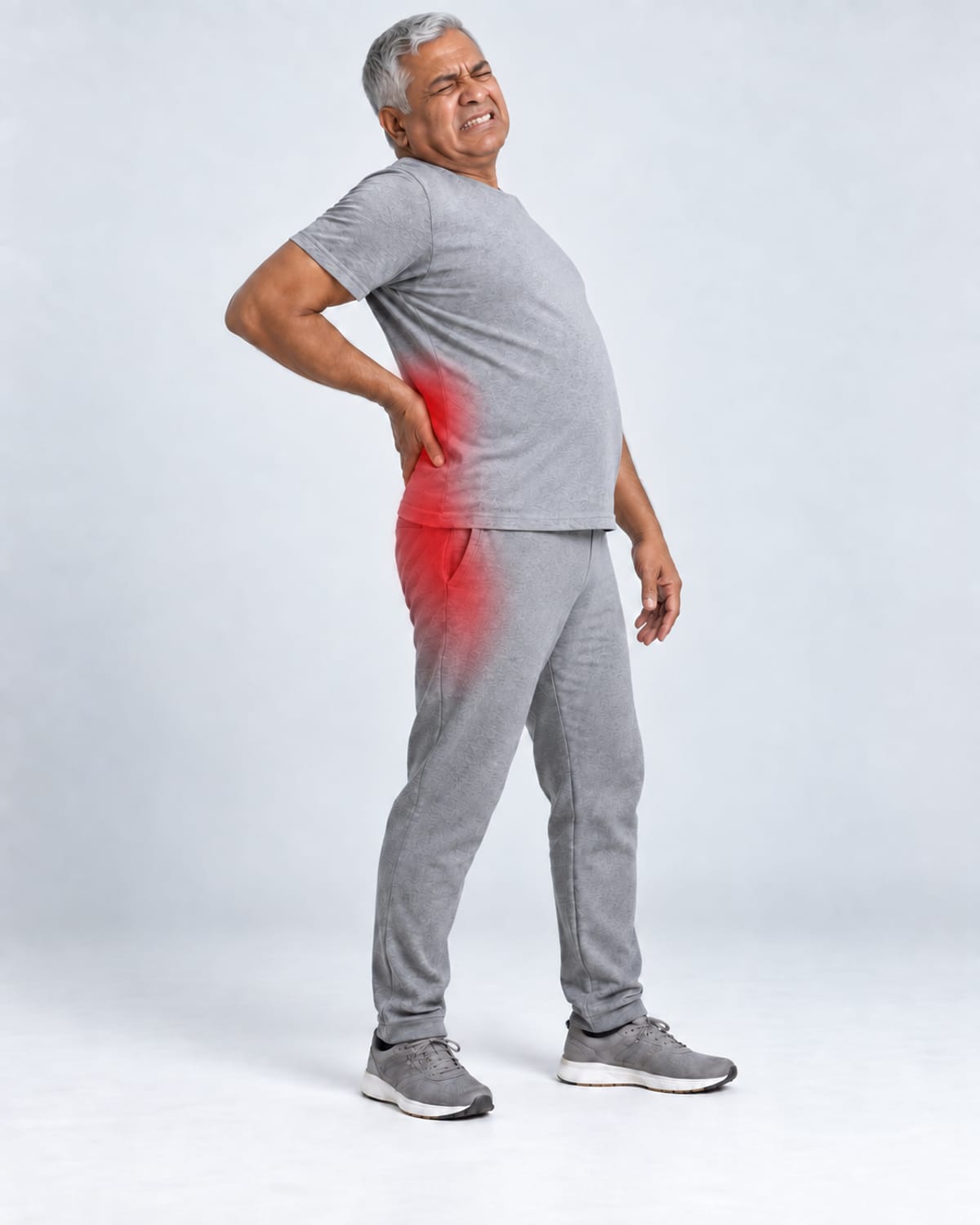
Dr Yas’s 7-Step Check: how we find the cause
The steps are the same ones used for a painful knee replacement, in an order that suits the hip. Infection comes first because it hides. “Is it the hip at all?” comes second because it is so often the answer. Every patient gets the results written down.
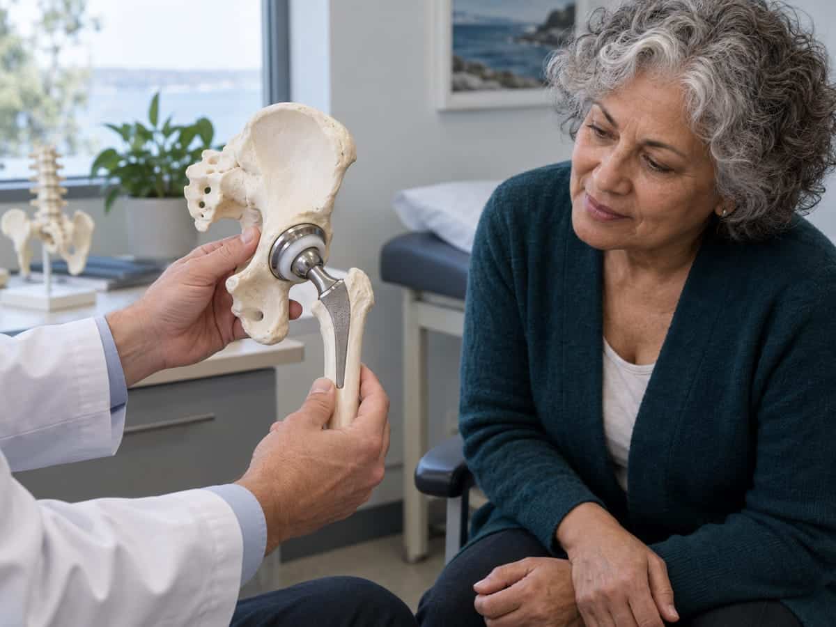
Listen, then examine.
Where exactly does it hurt — groin, buttock, side, thigh? When did it start? Has it hurt since the operation, or did a good hip turn bad? What makes it worse: standing up, walking far, lying on it, lifting the leg into the car? Then an examination of the hip, the back, the pelvis and the way you walk.
Rule out infection.
Two blood tests, CRP and ESR, which rise with inflammation. A normal result does not clear you: in one study of 215 proven joint infections, the CRP was normal in more than a third, almost all of them slow-growing bugs.9 So if the hip has been painful for months, the next step is to look inside the joint. Fluid drawn off with a needle, under X-ray or ultrasound guidance, is a start — but the slow-growing bugs live in the lining of the joint, not the fluid, so often what is needed is a tissue sample: several small pieces of the lining, taken through a keyhole procedure and grown in the lab for two weeks.10,11,23

Is it the hip at all?
The back and the pelvis are examined and, if the story fits, imaged. A pinched nerve, a narrowed spinal canal or a sacroiliac joint can all masquerade as the hip. Sometimes an injection of local anaesthetic into the hip joint settles it: if the pain goes, it was the hip; if it doesn’t, it wasn’t.6,8
Is the implant loose, worn or in the wrong place?
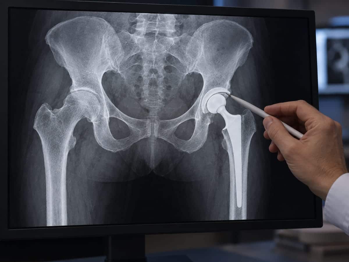
Standing X-rays of the pelvis and the whole leg. A CT scan measures where the cup and stem sit and how far the cup sticks out at the front. A bone scan or SPECT-CT shows whether the bone around the implant is working too hard, which is what loosening looks like.12 Leg length is measured on the scan.
Is it the soft tissue around the hip?
Two problems account for most of these. Pain on the outside of the hip, worse lying on it, is usually the gluteal tendons and the bursa over the point of the hip — the single most common finding in the study above.6,13 A catch or ache at the front of the hip when you lift the leg is usually the psoas tendon rubbing on the front edge of the cup.14,15 Both are confirmed with ultrasound and, if needed, a guided injection. Both are dealt with below.
Is it the implant itself?
Older implants used a type of plastic that wore faster and could make the bone around it thin, and some used metal-on-metal bearings that can irritate the tissues.16,17 Where a metal ball meets a metal stem, corrosion can do the same.6 Blood metal levels and a special MRI answer this.
Is the hip unstable or catching on itself?
A hip that clunks, feels loose, or has come out of joint needs the position of the parts measured on CT. Bony or soft-tissue impingement — the implant or the bone catching at the end of movement — is checked the same way.
The rare ones.
Nerve injury, heterotopic bone (extra bone forming in the muscle), complex regional pain syndrome, and metal allergy. Real, uncommon, and considered last.6
The rule at the end of the seven steps: name the cause first.
If the steps find nothing, the next step is a second look and a discussion with colleagues, not a guess.
Is it the hip joint, or the tendons on the side?
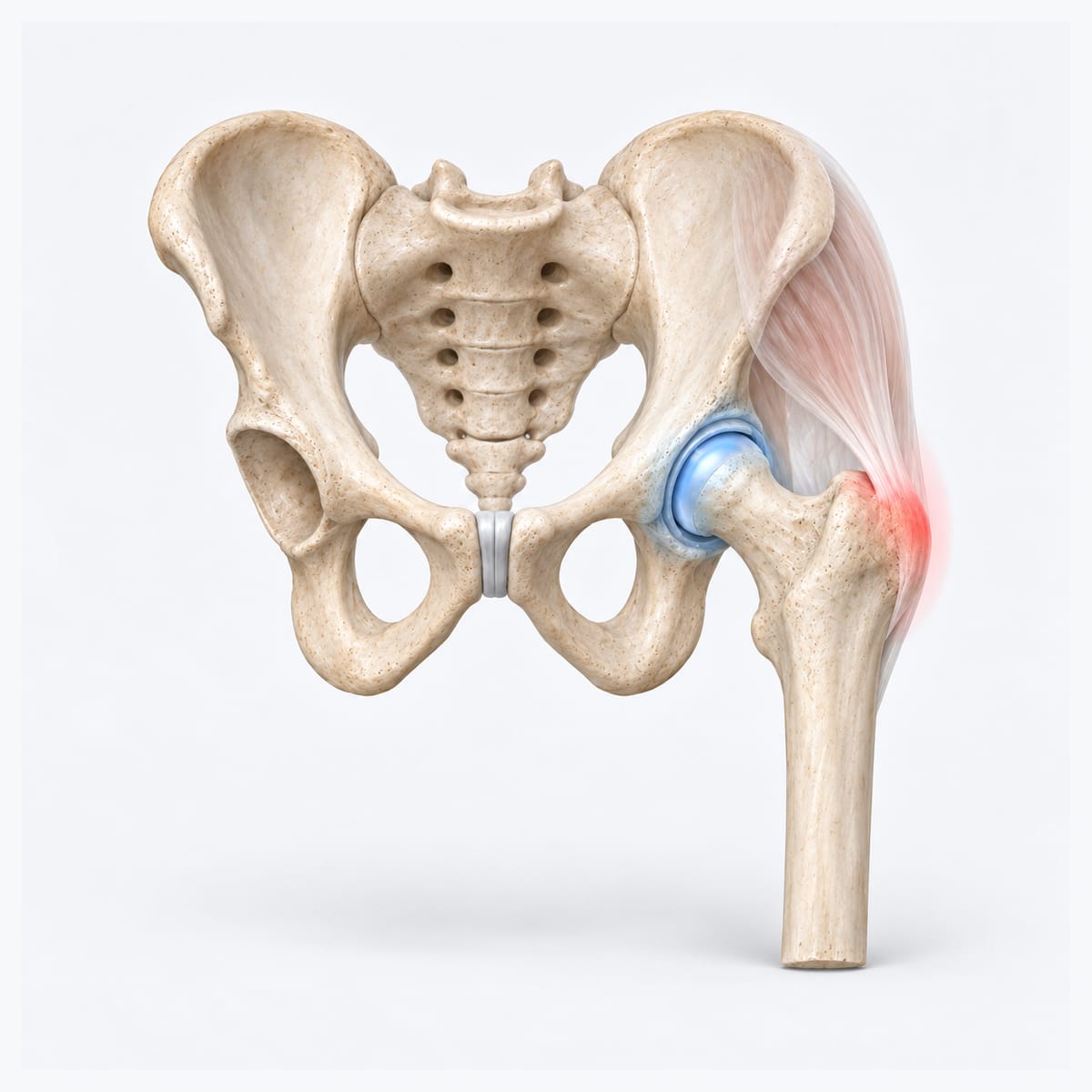
This is the most common mix-up Dr Yas sees, and it matters, because the two problems need completely different treatment. The hip joint is a ball and socket deep in the groin. The tendons are on the outside, over the bony point of the hip. They are a hand’s width apart, and they behave differently.
| The hip joint (arthritis, or a hip replacement problem) | The tendons on the side (gluteal tendons, bursa, ITB) | |
|---|---|---|
| Where it hurts | Deep in the groin, sometimes the front of the thigh or the knee | On the bony point on the outside of the hip, sometimes down the outside of the thigh |
| How people point to it | A hand cupped around the front and side of the hip — the “C sign” | One finger on the point of the hip |
| What makes it worse | Putting on socks and shoes, getting in and out of a car, turning over in bed, the first steps after sitting | Lying on that side at night, standing on one leg, walking up hills or stairs, sitting with legs crossed |
| Stiffness | Yes — the joint loses movement; shoes and socks get hard | No — the joint moves freely; it is sore, not stiff |
| What a hip replacement does | Replaces the worn joint | Nothing — the tendons are outside the joint |
| What helps | Depends on the cause found in the 7-Step Check | Strengthening physio (not stretching), a guided injection, and for the stubborn ones a keyhole release |
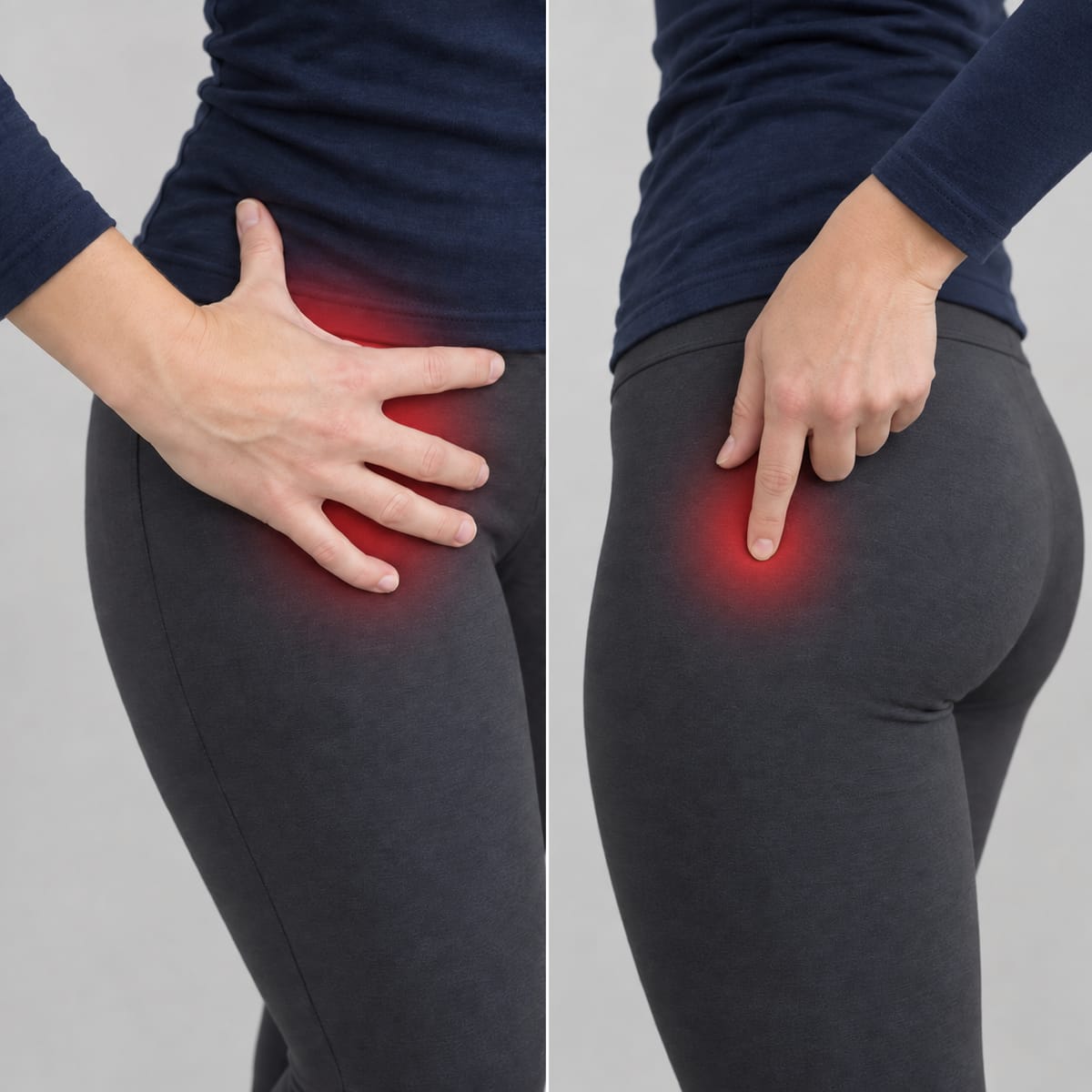
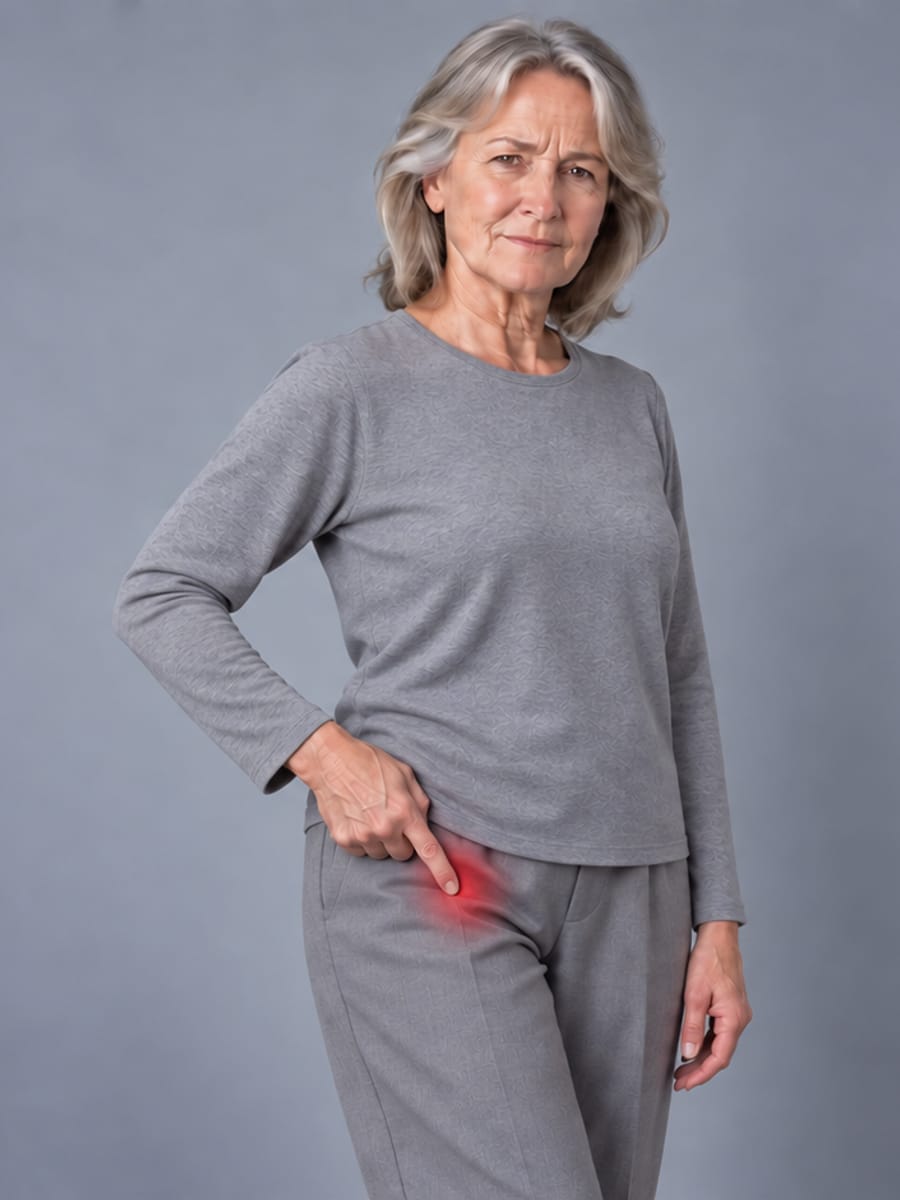
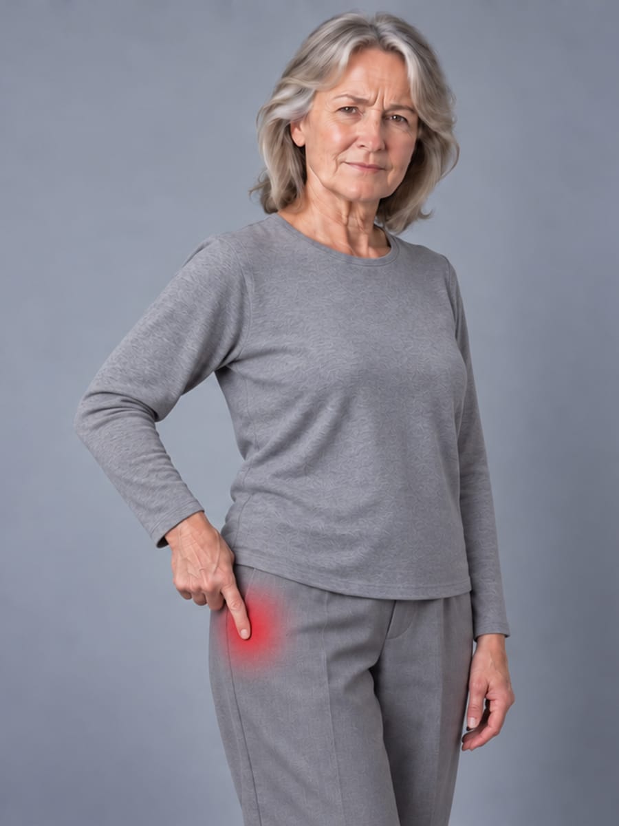
Both can exist at the same time, and one can hide the other. A person with arthritis often also has sore tendons from limping, and a person with a good hip replacement can still have tendon pain that was there before the surgery. Sorting out which is which — by examination, and sometimes by an ultrasound or an injection test — is one of the most useful things a first visit can do.6,13,22

Dr Yas’s most-watched video is about exactly this: “Pain on the SIDE of your hip is one of the most common things I see — and one of the most misunderstood.”
Two tendon problems that are easy to miss after a hip replacement
Both of these are tendon problems, not implant problems, and both are more common — and harder to live with — than they sound. They are easy to under-rate: the X-ray of the replacement looks perfect, so the pain gets labelled “muscular” and left to run its course. Physiotherapy is the right first step, but when it is not enough the problem still has a name and a fix. A hip surgeon who looks for these tendon problems can pick them up at the first visit and treat them alongside the replacement — often without touching the implant at all — so your new hip can do its job.
Pain on the outside of the hip
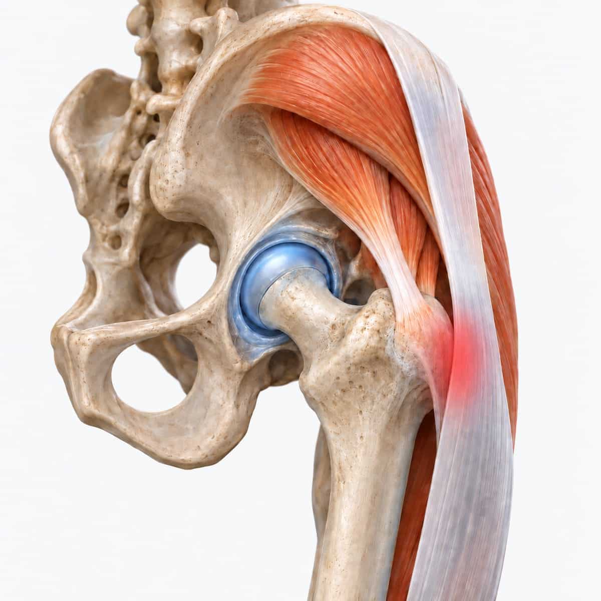
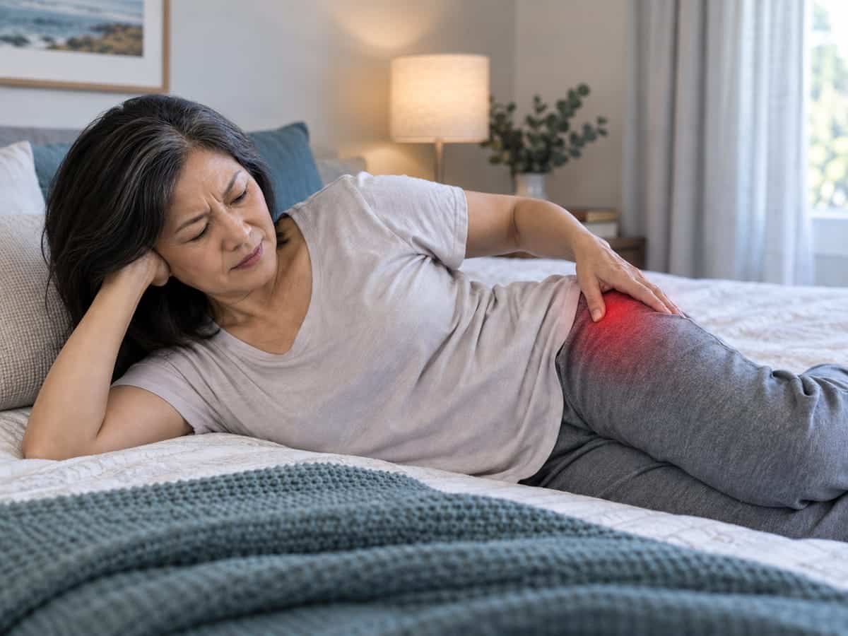
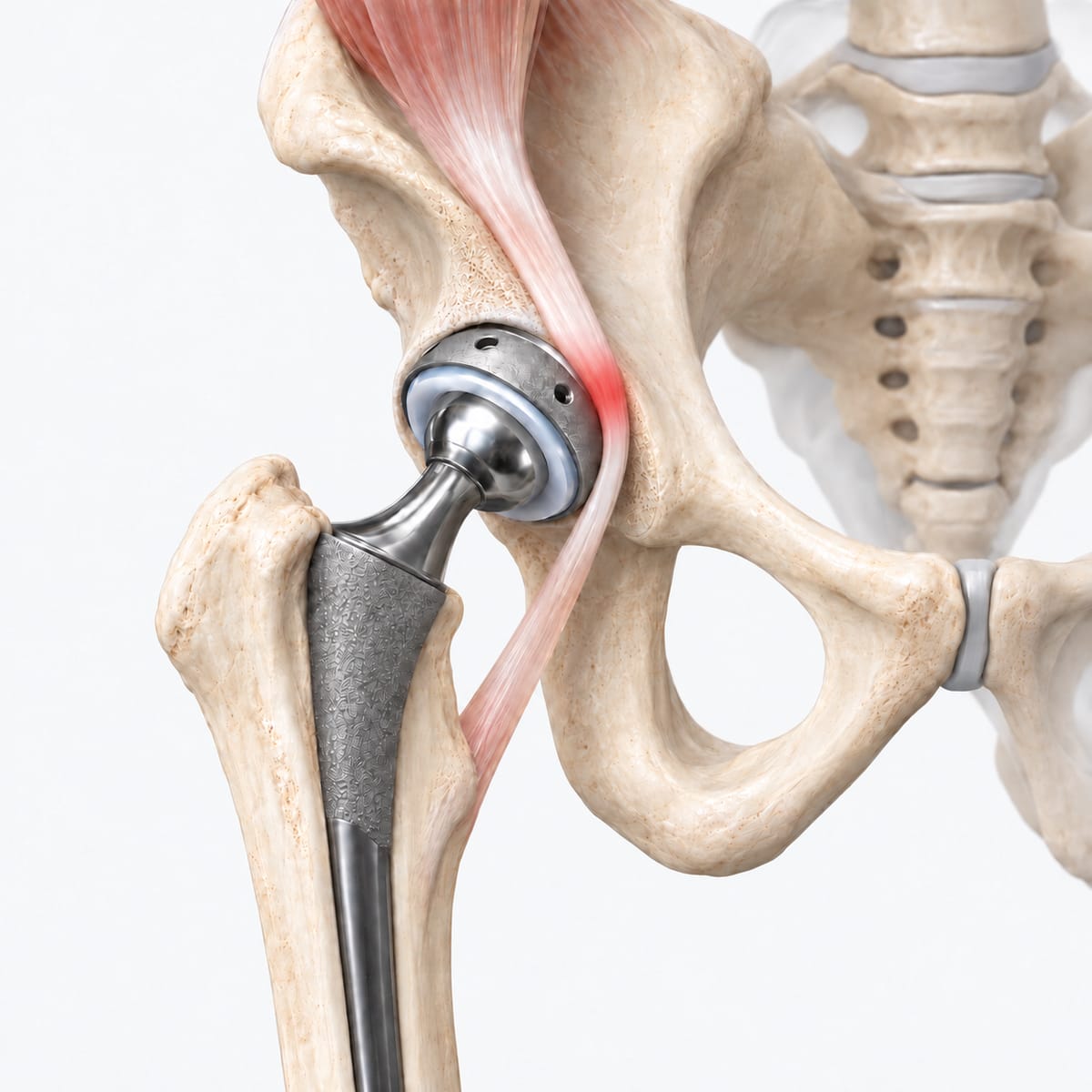
The point you feel on the side of your hip is the greater trochanter. Over it run the gluteal tendons — the tendons of the buttock muscles that hold your pelvis level when you walk — with a thin cushion (a bursa) and the long iliotibial band on top. When the tendons are worn or the band is tight, you get pain on the outside of the hip, a limp, and a hip you cannot lie on at night. Doctors call it gluteal tendinopathy, trochanteric bursitis or greater trochanteric pain syndrome. It is the same thing.
This is not arthritis, and a hip replacement does not fix it — the tendons sit outside the joint, so a new joint leaves them exactly as they were. After a hip replacement, about 1 in 20 people develop it, more often when the surgery went through the side of the hip.13 It was the single commonest finding in hips that still hurt at six months.6 Because the X-ray looks normal, it is easy to under-rate, and people can live with it for years longer than they need to.
Most cases settle with the right physiotherapy — strengthening, not stretching — and, when needed, a guided injection. For the ones that don’t, there is a keyhole (endoscopic) option: releasing the tight band and clearing the inflamed bursa through two small cuts, usually as day surgery. Dr Yas performs this procedure. In published series, 7 to 10 out of every 10 patients get lasting relief.18,19 A torn tendon can be repaired at the same time. He treats the tendons and the replacement as one problem, not two: a sore tendon can be sorted out while the replacement is left alone, and if the hip itself also needs attention, both can be dealt with at the same time.
Read more: Pain on the outside of the hip →
A catch at the front of the hip
The psoas is the big muscle that lifts your leg. Its tendon runs directly over the front edge of the hip socket. If the cup of a hip replacement sits a few millimetres proud at the front, the tendon rubs on it every time you lift the leg — getting out of a chair or a car, climbing stairs. The pain is at the front of the hip or groin, and the X-ray usually looks fine. That is exactly why it is so easy to miss: nothing shows on the film, and the catch only appears when the leg lifts.
An ultrasound-guided injection around the tendon is both the test and the first treatment. If the relief is real but short-lived, the tendon can be released where it catches, through keyhole surgery — a procedure Dr Yas performs. In the largest series, about 9 in 10 people had their pain settle, with a low complication rate and most home the same day.15 If the cup sits well forward — more than about 8 millimetres proud — releasing the tendon is not enough, and the cup itself is repositioned.14 Dr Yas looks for this at the first visit, because it is one of the most fixable causes of a painful hip replacement: the tendon can be treated on its own, and if the cup also needs attention, both are dealt with at the same time.


When the implant itself is the reason
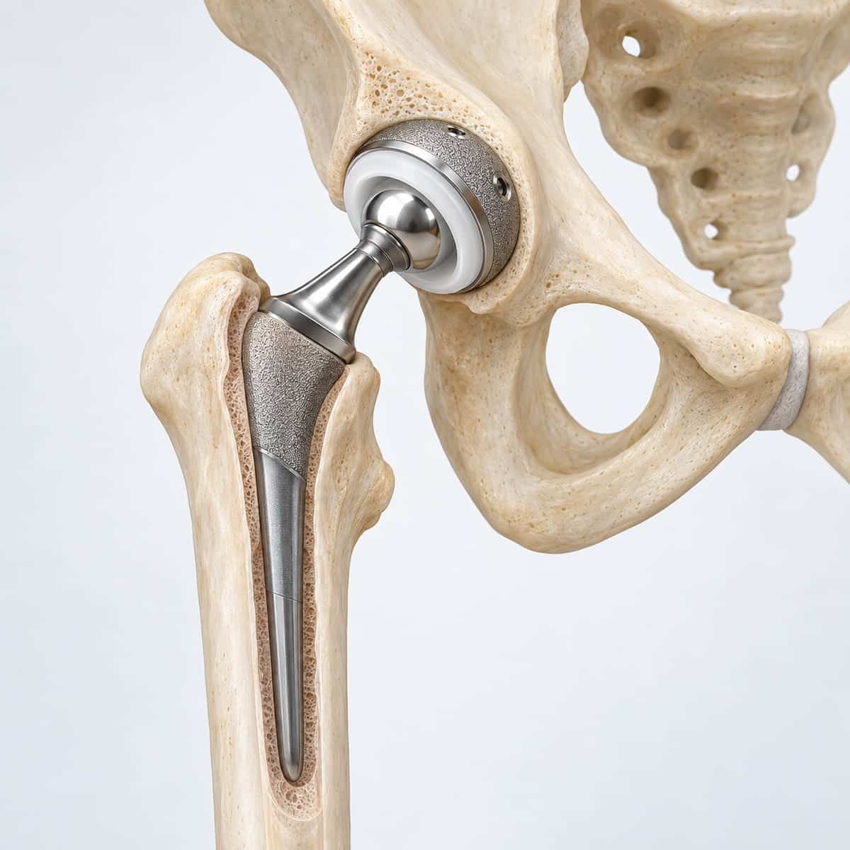
The plastic liner in a hip replacement has changed. The older type wore faster, and the tiny particles it shed made the bone around the implant thin — a process called osteolysis — which can loosen the cup or stem years later. A tougher, “highly cross-linked” plastic arrived in 1998 and took most of the 2000s to become standard, so a hip put in before about 2010 may still have the older kind.16,24 The newer plastic cut the problem from roughly one hip in four to one in twenty.16 Some hips put in during the 2000s used a metal ball in a metal cup. A proportion of these release metal into the tissues, which can cause pain, swelling and damage to the muscles; the UK registry found they failed much more often, and the bigger the ball, the sooner.17 Where a metal ball sits on a metal stem, corrosion at the join can do the same, even with a plastic cup.6
If you have an older hip that has started to hurt, this step matters: a blood test for metal ions, a specific type of MRI, and a plan before anything is damaged further.
What usually helps, once the cause is known
| If the cause is… | The treatment is usually… |
|---|---|
| Infection | Clearing the infection — antibiotics with a washout, or replacing the implant in one or two stages |
| The back | Treating the back — the hip is left alone |
| Gluteal tendons / bursa on the outside | Physio and a guided injection; keyhole release of the band if it doesn’t settle |
| Psoas tendon catching on the cup | Guided injection; keyhole tendon release; cup repositioned if it sits too far forward |
| A loose, worn or badly placed implant | Revision surgery — replacing the worn or loose part with implants designed for the job |
| An old bearing or corrosion | Exchanging the bearing, sometimes the cup or stem, before the tissues are damaged |
| Instability or impingement | Repositioning the parts, or a larger bearing that resists dislocation |
| Nothing found | A second look, a discussion with colleagues, and an honest answer |

You don’t need to bring anything
Just come as you are. Dr Yas tracks down what is needed himself — the operation report and the details of the implant from the hospital where the surgery was done, including the public hospitals, and any X-rays and scans you have had. If blood tests or new scans are needed, he arranges them. If you happen to have letters or results, bring them by all means, but nothing is expected of you. A GP referral helps with Medicare but is not required.

Common questions
How do I know if it’s my back or my hip?
I can’t lie on that side. Is the replacement failing?
The X-ray looks perfect. So why does it hurt?
My blood tests are normal. Can it still be infected?
Will I need another operation?
Will my surgeon mind if I get a second opinion?
Do I need a GP referral?
Ask Dr Yas about your hip
Dr Yas wants you to ask questions. Send a few details and what has been happening, and the rooms will be in touch within one business day. A GP referral helps but is not required.
- A proper assessment — your history, an examination of the hip and the back, and your scans reviewed with you
- The seven steps, in order, with the results written down
- A clear plan — and an honest “we don’t know yet” if that is where the tests land
Have a question about your hip?
Ask about what has been happening, the tests, or whether a second look makes sense for you. It takes two minutes.
Do you have private health insurance?
Send your question
Write as much or as little as you like.
Thank you
The rooms can talk you through your options and what a consultation involves.
Contact Dr Yas- Vaotuua D, O’Connor P, et al. The effect of surgeon volume on revision for periprosthetic joint infection: an analysis of 602,919 primary total knee arthroplasties (AOANJRR). J Bone Joint Surg Am 2023;105:1663–75.
- British Orthopaedic Association / BASK. BOAST: Revision total knee replacement surgical practice guidelines. August 2020.
- The effect of minimum volume recommendations on surgeon activity for first revision total knee replacement: an analysis of 2009–2019 UK National Joint Registry data. The Knee 2025.
- Beswick AD, Wylde V, et al. What proportion of patients report long-term pain after total hip or knee replacement for osteoarthritis? A systematic review of prospective studies in unselected patients. BMJ Open 2012;2:e000435.
- Lanting BA, MacDonald SJ. The painful total hip replacement: diagnosis and deliverance. Bone Joint J 2013;95-B(11 Suppl A):70–3.
- Erivan R, Villatte G, Ollivier M, Paprosky WG. Painful hip arthroplasty: what should we find? Diagnostic approach and results. J Arthroplasty 2019;34:1802–7.
- Lenguerrand E, Wylde V, et al. Trajectories of pain and function after primary hip and knee arthroplasty: the ADAPT cohort study. PLoS One 2016;11:e0149306.
- Devin CJ, McCullough KA, et al. Hip-spine syndrome. J Am Acad Orthop Surg 2012;20:434–42.
- Akgün D, Müller M, et al. The serum level of C-reactive protein alone cannot be used for the diagnosis of prosthetic joint infections, especially in those caused by organisms of low virulence. Bone Joint J 2018;100-B:1482–6.
- Parvizi J, Tan TL, et al. The 2018 definition of periprosthetic hip and knee infection: an evidence-based and validated criteria. J Arthroplasty 2018;33:1309–14.
- Shohat N, Bauer T, et al. Hip and knee section: what is the definition of a periprosthetic joint infection of the knee and the hip? J Arthroplasty 2019;34:S325–7.
- Van den Wyngaert T, Paycha F, et al. SPECT/CT in postoperative painful hip arthroplasty. Semin Nucl Med 2018;48:425–38.
- Iorio R, Healy WL, et al. Lateral trochanteric pain following primary total hip arthroplasty. J Arthroplasty 2006;21:233–6.
- Chalmers BP, Sculco PK, Sierra RJ, Trousdale RT, Berry DJ. Iliopsoas impingement after primary total hip arthroplasty: operative and nonoperative treatment outcomes. J Bone Joint Surg Am 2017;99:557–64.
- Guicherd W, Bonin N, et al. Endoscopic or arthroscopic iliopsoas tenotomy for iliopsoas impingement following total hip replacement: a prospective multicentre 64-case series. Orthop Traumatol Surg Res 2017;103(8S):S207–14.
- Prock-Gibbs H, Pumilia CA, et al. Incidence of osteolysis and aseptic loosening following metal-on-highly cross-linked polyethylene versus conventional polyethylene total hip arthroplasty: a systematic review at medium to long-term follow-up. J Bone Joint Surg Am 2021;103:728–40.
- Smith AJ, Dieppe P, et al. Failure rates of stemmed metal-on-metal hip replacements: analysis of data from the National Joint Registry of England and Wales. Lancet 2012;379:1199–204.
- Giai Via R, Elzeiny A, et al. Endoscopic management of greater trochanteric pain syndrome: a systematic review. Eur J Orthop Surg Traumatol 2024;34:3385–94.
- Walker-Santiago R, Wojnowski NM, et al. Platelet-rich plasma versus surgery for the management of recalcitrant greater trochanteric pain syndrome: a systematic review. Arthroscopy 2020;36:875–88.
- Dora C, Houweling M, et al. Iliopsoas impingement after total hip replacement: the results of non-operative management, tenotomy or acetabular revision. J Bone Joint Surg Br 2007;89-B:1031–5.
- Ueno T, Kabata T, et al. Risk factors and cup protrusion thresholds for symptomatic iliopsoas impingement after primary total hip arthroplasty. J Arthroplasty 2018;33:3288–96.
- Chamberlain R. Hip pain in adults: evaluation and differential diagnosis. Am Fam Physician 2021;103:81–9.
- Fink B, Makowiak C, et al. The value of synovial biopsy, joint aspiration and C-reactive protein in the diagnosis of late peri-prosthetic infection of total knee replacements. J Bone Joint Surg Br 2008;90-B:874–8.
- de Steiger R, Lorimer M, Graves SE. Cross-linked polyethylene for total hip arthroplasty markedly reduces revision surgery at 16 years (AOANJRR 1999–2016). J Bone Joint Surg Am 2018;100:1281–8.